September 2024: CLSM Image with REMBI Annotations on OMERO
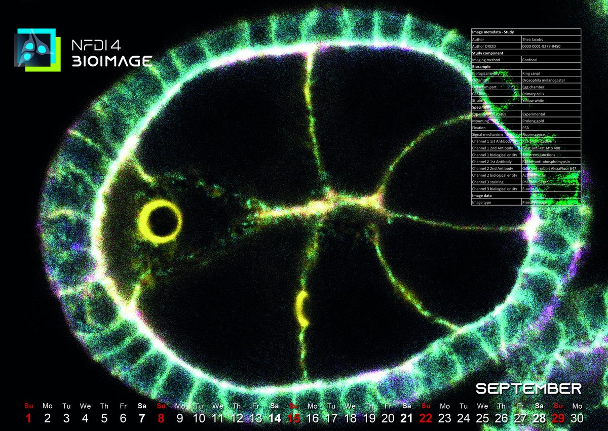
This month, our featured calendar image showcases an early-stage oocyte from a Drosophila melanogaster egg chamber. Thea Jacobs from the Luschnig lab, which focuses on epithelial morphogenesis and paracellular barrier formation, in the Institute of Integrative Cell Biology and Physiology of University Münster, skillfully captured it using the advanced capabilities of a Leica SP8 Confocal Laser Scanning Microscope (CLSM). The specimen, representing the wildtype of this model organism provides critical insights into developmental biology.
The image itself strikingly details the actin ring canals across the nurse cells, pivotal for the exchange of cytoplasm with the developing oocyte. These ring canals are instrumental in the development of the oocyte, ensuring proper distribution and communication among the cells within the egg chamber. Highlighted in this microscopic capture are the surrounding structures as well—the germline cells encapsulated by a monolayer epithelium, which provides the necessary cellular interaction and support during the egg's development.
This month, the highlight is not just the scientific insight the image provides or the sophisticated imaging techniques used, but predominantly its public accessibility. Emphasizing the commitment to transparency and openness in scientific research, this detailed image data is made publicly available via the OMERO instance of the University of Münster. Interested parties can delve into the data themselves here.
This accessibility allows researchers, educators, and the curious public alike to explore not only the intricate biological structures depicted but also to examine the comprehensive metadata adhering to the REMBI standards, aiding in the broader dissemination and understanding of this developmental biology data. Consequently, it ensures it meets the criteria of the FAIR principles — Findable, Accessible, Interoperable, and Reusable.
Images like these are more than just pieces of scientific art; they carry with them the capability to enhance our understanding of biological processes. This image was not merely captured; it was curated with a profound respect for both scientific pursuit and data management, echoing the mission of initiatives like NFDI4BIOIMAGE that advocate for improved research data management practices in the field of bioimaging.
This image stands out as a testament to the power of combining high-resolution microscopy with stringent data management protocols. It serves as a beacon for ongoing and future projects to adhere to high standards of documentation and compliance with FAIR principles, ensuring that every piece of data generated can be maximally utilized for scientific advancement. Join us in appreciating this remarkable depiction of early life at the microscopic level.